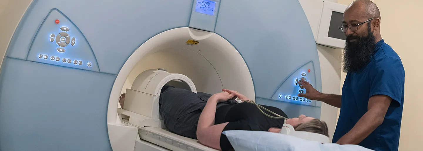
Imaging plays a critical role in diagnosing orthopaedic injuries and conditions, often serving as the first step in understanding what’s causing your pain or limited mobility. At Great Basin Orthopaedics, we offer on-site imaging services—including X-ray and MRI—to streamline your care, reduce wait times, and ensure your provider has immediate access to the information needed to guide your treatment.
Having imaging available in the same location as your clinical visit allows for faster answers, more accurate diagnoses, and a more seamless patient experience from evaluation through recovery.
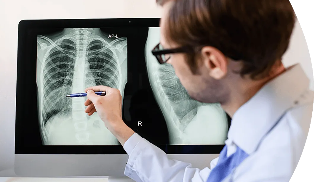
X-ray imaging is typically the first tool used to evaluate bone-related injuries. It's fast, widely available, and highly effective at identifying structural issues within the body.
What Is an X-Ray?
X-rays are a type of radiation called electromagnetic waves that create a picture of the inside of your body.
Is It Safe?
Generally, the amount of radiation you're exposed to during an X-ray is the equivalent to a few days of exposure to natural radiation from the environment.
When Are X-Rays Used?
X-rays are most commonly used to detect bone fractures, dislocations, and other structural abnormalities.
What to Expect
During your X-ray, you may be asked to wear a lead apron to protect certain areas of your body. The technician will step behind a shield or into another room while the images are taken. Depending on the area being examined, you may be asked to sit, stand, or lie down. Multiple images may be needed, but the process itself typically takes only a few minutes.
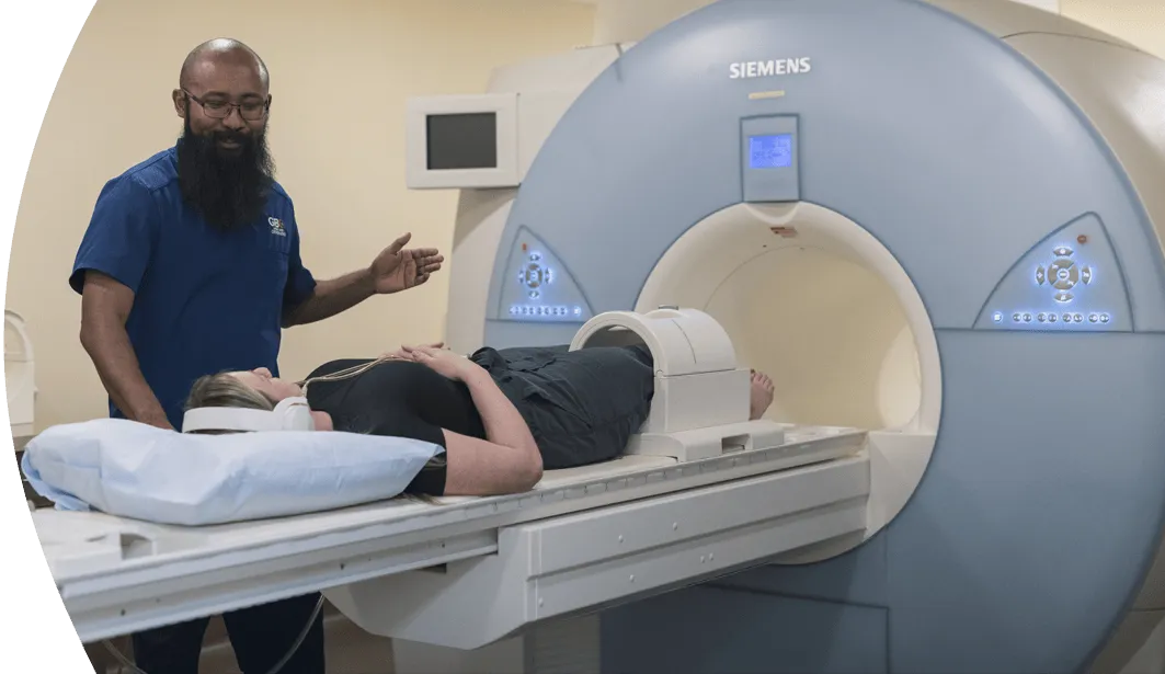
MRI is a more advanced imaging technique used when a more detailed view is needed, especially for soft tissues that don't appear clearly on X-rays.
What Is an MRI?
Magnetic resonance imaging (MRI) uses a powerful magnet, radio waves, and computer technology to produce detailed, cross-sectional images of the body's internal structures. It is noninvasive and does not use radiation.
When Are MRIs Used?
MRI is often used to identify conditions not visible on X-rays, such as small fractures, ligament tears, cartilage damage, and tendon injuries. Its ability to highlight contrasts in soft tissue makes it especially useful for evaluating joints, cartilage, ligaments, and tendons. It is also helpful in detecting infections, inflammation, and other soft tissue abnormalities, and can assist in ruling out more serious conditions.
What to Expect
MRI uses a magnet to collect images, so items made of metal are not allowed near it (remove all jewelry and alert the technician of any metal in your body, like pacemakers or surgical hardware). During the MRI scan, you lie on a table that slides inside a tunnel-shaped machine. Scans typically take between 20 and 60 minutes, during which time you will need to remain still. While MRI machines make a lot of noise (you'll be given ear protection), the scan is painless. Some scans require only part of the body to be inserted into the machine, and others require the entire body which may be uncomfortable for those who are prone to claustrophobia.